Methods for determining urea in blood serum. Clinical significance of urea determination. Determination of urea in blood serum and urine with diacetyl monooxime
Urea or urea or carbonic acid diamide is what ultimately remains of proteins after they break down.
Many people confuse urea with uric acid (the result of purine metabolism) and, it should be noted, they have something related, for example, they both belong to the group of residual nitrogen components, but in clinical laboratory diagnostics these indicators carry different concepts and cannot be considered as one whole.
Urea and its norm
The level of urea in the blood can fluctuate down or up due to completely physiological circumstances. For example, it is influenced by nutrition, physical activity, and in women the level of urea in the blood is slightly lower than in men. If there is a lack of protein in the diet, urea will be reduced, and if there is too much, then it will increase.

A diet depleted in chlorine, for example, refusal of table salt, will increase urea - this is an adaptive mechanism activated by the body (after all, is it necessary to somehow maintain colloid-osmotic pressure?).
Pregnancy does not obey generally accepted laws, we are not talking about one specific life, therefore many biochemical indicators, adapting to this crucial period, behave differently, urea, for example, decreases, but this is normal. Women with a complicated medical history (pyelonephritis, glomerulonephritis, kidney stones) are under special control, since there is a risk of developing renal failure and uremic syndrome.
The normal level of urea in the blood of an adult healthy person is within the range 2.5 – 8.3 mmol/liter. In women, this figure is usually lower, but they do not have a separate norm. Urea excretion in urine is 20.0 – 35.0 g/day (333.6 – 587.7 mmol/day).
"Urine in the Blood"
A strongly increased concentration of urea in the blood, which occurs as a result of acute and chronic renal failure, is well known to specialists in various fields and is called uremic syndrome(“urinary bleeding”). In addition to urea, with uremia there is an accumulation of ammonia, uric acid and many other protein breakdown products, which poison the body and can quickly lead to death.

Uremia, caused by the accumulation of nitrogenous waste in the body, is accompanied by symptoms of severe intoxication, although it all starts with the usual manifestations of fatigue:
- Brokenness;
- General weakness;
- Fast fatiguability;
- Headache.
Such seemingly harmless symptoms soon to join:
- Disturbance of homeostasis with disruption of the activity of many organs, which can be suspected when nausea, vomiting, and diarrhea occur;
- Lack of urine (anuria);
- Severe liver dysfunction;
- Visual impairment;
- Tendency to bleeding;
- Changes in the skin (uremic “powder”).
The nitrogenous components that are not lost in the urine are looking for a way out. They seep through the skin (uremic (“frost”), serous and mucous membranes, causing their damage. Particular suffering falls on the digestive organs, urogenital tract, eyes, but the skin is most visible, so people say: “urine went through the skin” It is difficult to treat such conditions, but in cases of acute renal failure, despite the very rapid development of events, With timely and adequate treatment (hemodialysis), complete restoration of the body is possible.
In the chronic form of uremic syndrome, in addition to all the changes in the kidneys, it quickly develops with very high blood pressure numbers, blood circulation in all organs is disrupted, and develops. A person’s life can be extended, mainly due to hemodialysis (even up to 20 years), but, in the end, the terminal stage of the disease occurs (pneumonia, sepsis, uremic coma), which, as a rule, leaves no chance.
It is possible to save the patient (of course, to the terminal stage of uremic syndrome!) will be able to get a donor kidney, which, as you know, is not lying around on the road, so patients have been on waiting lists for years. Relatives, unfortunately, are not always suitable; moreover, they themselves often have a similar pathology (after all, they are relatives).
Individual abilities of urea
Urea itself, unlike some other slags (ammonia, cyanate, acetone, phenols), is not toxic, but has its own abilities. It can easily penetrate through plasma cell membranes into parenchymal organs (liver, kidneys, spleen) and, having osmotic activity, pulls water with it, which leads to swelling of cells (hypehydration), which lose the ability to function normally.
Due to the fact that urea penetrates cells well, it passes through the membranes of the kidney filter with the same success, and therefore is excreted remarkably in the urine. There is the same amount of carbamide in the glomerular filtrate as in plasma, but moving along the tubules, it can give off water and be absorbed itself (tubular reabsorption). At the same time, the higher the rate of urine flow, the less the urea content will change (it simply does not have time to return). It is clear that in case of impaired renal function (renal failure), a large amount of urea from the water will return back to the body and be added to what is in the plasma - this is the increased level of urea in the blood.
It may follow from this that low urea in the blood occurs if a person’s diet contains few protein foods, and urine in the kidney moves at high speed and the urea does not have time to return.
It's not just the kidneys that are to blame
An increased concentration of urea in the blood, as noted earlier, is observed with excessive consumption of foods rich in proteins or a diet depleted in chlorine. In addition, an increase in the level of urea can cause pathological conditions associated either with increased formation of urea, or with the retention of nitrogenous waste for some reason.
- Increased protein breakdown and, accordingly, increased urea biosynthesis (productive azotemia) cause many serious human diseases:
- Hematological diseases (leukemia, malignant form, hemolytic jaundice).
- Severe infections, including intestinal ones (dysentery, typhoid fever, cholera).
- Intestinal diseases (obstruction, peritonitis, thrombosis).
- Burn disease.
Neoplasms of the prostate gland. Retention of nitrogenous wastes (urea, in particular) and their slow excretion in the urine as a result of impaired functional abilities of the excretory system (retention renal azotemia) or as a result of other reasons (retention extrarenal azotemia)
- often accompany various renal and other pathologies:
- Pyelo- and glomerulonephritis;
- Polycystic kidney disease;
- Nephrosis;
- Acute and chronic renal failure (ARF and CRF);
- Sublimate poisoning;
- Tumors of the urinary tract;
- Urolithiasis (UCD);
- Reflex anuria;
- Decompensated (impaired renal hemodynamics);
- Gastrointestinal bleeding;
Slow excretion of urea in the urine is observed in cases of renal dysfunction, nephritis, uremic syndrome (nephropathy of pregnancy), the use of anabolic steroids, severe liver damage (in this case, it simply stops being produced by the liver parenchyma, so its content in the blood does not increase).
Low in blood, high in urine and other options
The causes of low urea in the blood were also slightly touched upon above (lack of nutrition or complete starvation, pregnancy). However in some cases, urea is reduced due to very serious circumstances:
- Extremely severe liver damage (parenchymal jaundice, acute dystrophy, decompensated cirrhosis), because urea biosynthesis occurs in this organ.
- Poisoning with hepatotropic poisons (arsenic, phosphorus).
- Reduced metabolic breakdown of proteins.
- After hemodialysis and glucose administration.
Increased level of urea in urine, that is, its increased secretion by the kidneys can become a sign of disease or oversaturation of the body with protein:
- Pernicious anemia (nitrogen imbalance);
- The use of certain medications (quinine, salicylates);
- Feverish conditions;
- Postoperative period;
- Increased thyroid function;
- Overdose of L-thyroxine;
- Administration of 11-OX (11-hydroxycorticosteroids).
As for the hyperprotein diet. If a person intensively consumes protein-rich foods, then it is quite natural that a healthy body will intensively remove the products of protein catabolism (the level of urea in the urine is increased), trying to prevent any significant changes in blood levels. Although if such a diet becomes the meaning of life, then urea in the blood will eventually begin to increase.
Reduce blood urea (in conclusion)
A diet will help reduce urea in the blood, if its increase is caused by not very serious reasons. Maybe you shouldn’t always fill your breakfasts, lunches and dinners with protein foods? Maybe, Sometimes it’s better to add more vegetables and fruits to the table, and they will solve the problem.
Well and if urea in the blood is low, then, along with your favorite foods of plant origin, you should think about protein foods, to allow the body to function normally.


Breakdown of proteins in the body accompanied by the release of urea. At the same time, nitrogen concentrated in urea is removed from the body along with it. An increased level of urea in the blood is a sign of renal failure, resulting, for example, in uremia. That is why the determination of urea in blood serum becomes important.
Special research activities on blood serum are carried out in order to establish possible renal pathology, as well as the degree of its development. Timely analysis is an opportunity to reduce treatment time.
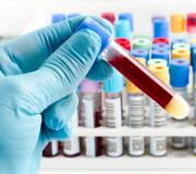
Analysis is possible in specialized medical laboratories that have the appropriate license to carry out such work, as well as technical equipment and chemical reagents. Blood serum tests for urea concentration can be carried out using several methods, divided into the following possible groups:
- Gasometric.
- Urease.
- Photometric straight lines.
Indications for research
 The standard level of urea concentration in the blood of an adult is 640-660 mg per liter. In athletes involved in weightlifting and bodybuilding, the presence of urea is slightly higher. This is due to the increased metabolism associated with consuming large amounts of protein. However, an increased urea value may be due to the following possible pathologies:
- Leukemia.
- Dysentery.
- Parenchymal jaundice.
- Pyelonephritis.
- Kidney failure (chronic form).
- Glomerulonephritis.
Principles of analysis
Regardless of the analysis method, the determination of uric acid has the following important features:
- Blood collection time is from 8 to 11 o'clock in the afternoon.
- The patient should not be hungry for more than 14 hours.
- Blood is taken from the patient's vein.
- Sufficient blood volume within 8 ml.
Research methods
To determine the exact concentration of uric acid in the blood serum, the patient may be offered the following possible options:
- Xanthhydrols.
- Hypochlorite.
- Diacetyl monooxime.
- Semi-quantitative methods using indicator paper.
- Methods using ion-selective electrodes.
- Enzymatic.
- Gasometric.
Xanthhydrol methods
The basic component involved in research is xanthhydrol(heterocyclic alcohol).
The principle of the method is to obtain a compound of xanthhydrol with urea and subsequent determination of the weight fraction of each substance.Hypochlorite methods
These methods for determining uric acid used infrequently, due to the high turbidity of the urea compound with laboratory reagents. The research involves sodium hypochlorite and phenol.
Diacetyl monooxime studies
 The determination of uric acid in the blood occurs through the Firon reaction, when the compound includes diacetyl monooxime and urea.
The result is a characteristic coloring of the elements used.
Semi-quantitative methods using indicator paper
 The advantage of the methods is the speed of obtaining data.
On average, the analysis takes no more than 10 minutes.
Test papers with applied reagents under the brands “Ureatest” and “Uranal” are widely distributed.
The principle of the analysis is almost the same as if a glucometer was used to measure cholesterol and sugar (this portable device successfully determines cholesterol levels in the human body).Methods using ion selective electrodes
When using the method, selective and another auxiliary electrodes are placed in the medium under study. The signal from a pair of electrodes is sent to a measuring device.
Enzymatic studies
Diagnosis through enzymatic studies involves hydrolysis of a certain volume of urea through urease.
The normal pH is in the range of 6.0-8.0.Gasometric methods
Another name for research is hypobromite analysis of urea concentration. The idea of the methods is to use the oxidation reaction and decomposition of urea through hypobromite. The reaction releases nitrogen and carbon dioxide. The last component is eliminated with a special solution, after which the amount of nitrogen is calculated.
The main groups of methods for determining urea content are divided into:
- Xanthhydrols: The heterocyclic alcohol xanthhydrol reacts with urea to form insoluble dixanthylurea, which is further determined gravimetrically, nephelometrically, colorimetrically or titrimetrically. The methods are accurate, but labor-intensive.
- Hypochlorite – B.A. Rashkovan’s method, which consists in the appearance of a characteristic color during the interaction of urea with sodium hypochlorite and phenol, is practically not used due to the different shades of the experimental and control samples, and the frequent appearance of turbidity when HCl is added.
- Diacetyl monooxime the methods are based on the Firon reaction - the interaction of urea and diacetyl monooxime with the formation of colored products, they are characterized by good reproducibility, high sensitivity, and high specificity.
- Semi-quantitative methods using indicator paper.
- Methods using ion selective electrodes.
- Enzymatic methods are based on the hydrolysis of urea by urease (optimum pH 6.0-8.0). The resulting ammonia is determined using various reactions (phenol hypochlorite, glutamate dehydrogenase, salicylate-hypochlorite).
- Gasometric(hypobromite), based on the oxidation and decomposition of urea with sodium hypobromite in an alkaline environment:
CO 2 (NH 2) + 3NaBrO → N 2 + CO 2 + 3NaBr + H 2 O
The released carbon dioxide is absorbed by the solution, leaving only nitrogen free, the volume of which is measured. The method is nonspecific (since hypobromite reacts not only with urea, but also with other substances containing amino groups), inaccurate, poorly reproducible and labor-intensive.
As unified Methods for determining urea have been approved: phenol hypochlorite, diacetyl monooxime and urease methods, and an express method using Ureatest indicator paper.
Determination of urea in blood serum and urine with diacetyl monooxime
The methods of this group are based on the Fearon reaction, which occurs in two stages. The first step is the hydrolysis of diacetyl monooxime to form diacetyl and hydroxylamine. In the second step, hydroxylamine reacts with urea to form a colored diazine derivative. For the oxidation of hydroxylamine, the following can be used: sodium persulfate, arsenic acid, perchloric acid, phenazone, cations. To intensify color and its stability, the following are used: thiosemicarbazide, phenylanthranilic acid, glucuronolactone, cations, tryptophan, nitrites.
Determination of urea by the urease method
Enzymatic methods are based on the hydrolysis of urea by urease in an incubation medium with pH = 6.0‑6.5 (EDTA buffer) or pH = 6.9‑7.0 (phosphate buffer) into carbon dioxide and ammonia. The resulting ammonia can be determined by a highly sensitive and specific reaction with phenol hypochlorite and the catalyst nitroprusside, by a salicylate-hypochlorite reaction, by a reaction with Nessler’s reagent (it is 10 times less sensitive compared to phenol hypochlorite, low specificity), by a dichloroisocyanurate reaction (the presence of protein interferes with the determination) .
Normal values
If it is necessary to compare the concentration of residual nitrogen with the nitrogen content of urea, the concentration of the latter should be divided by 2.14.
Influencing factors
- in vivo: increase - nephrotoxic drugs, corticosteroids, excess thyroxine; decrease - increase in the concentration of somatotropic hormone,
- in vitro: reduction (urease method) – sodium citrate.
Clinical and diagnostic value
Serum
The level of urea depends on the rate of its synthesis in the liver and excretion through the kidneys, as well as on the amount of protein catabolism.
Increased urea levels may occur with impaired renal function (acute and chronic diseases, urinary tract obstruction), impaired renal perfusion (congestive heart failure), depletion of body water (vomiting, diarrhea, increased diuresis or sweating), increased protein catabolism (acute myocardial infarction, stress, burns, yellow liver atrophy, gastrointestinal bleeding), with a high protein diet. In severe cases of acute renal failure, a 10-fold increase in urea levels was detected. Since the water-excreting function of the kidneys is restored faster than the concentration ability, normal excretion of urea in the urine occurs much later than the restoration of diuresis.
A decrease is observed with a low protein diet, with increased protein utilization in tissues (late pregnancy), severe liver diseases accompanied by impaired urea synthesis (parenchymal jaundice, liver cirrhosis).
Urine
An increase in the amount of urea in the urine is associated with hyperthyroidism, pernicious anemia, fever, phosphorus poisoning, observed with a high protein diet, and in the postoperative period.
The decrease is observed in patients with nephritis and other kidney diseases, uremia, parenchymal jaundice, cirrhosis or liver dystrophy, also in healthy growing children and on a low-protein diet.
Urea(also known as urea) is an organic compound having the formula CO(NH 2) 2, (carbonic acid diamide). Urea nitrogen makes up about 90% of the total nitrogen excreted.
Methods for determining urea content
The main groups of methods for determining urea content are divided into:
- Xanthhydrols: The heterocyclic alcohol xanthhydrol reacts with urea to form insoluble dixanthylurea, which is further determined gravimetrically, nephelometrically, colorimetrically or titrimetrically. The methods are accurate, but labor-intensive.
- Hypochlorite - B. A. Rashkovan’s method, which consists of the appearance of a characteristic color when urea interacts with sodium hypochlorite and phenol, is practically not used due to the different shade of experimental and control samples, and the frequent appearance of turbidity when adding HCl.
- Diacetyl monooxime methods are based on the Firon reaction - the interaction of urea and diacetyl monooxime with the formation of colored products; they are characterized by good reproducibility, high sensitivity, and high specificity.
- Semi-quantitative methods using indicator paper.
- Methods using ion-selective electrodes.
- Enzymatic methods are based on the hydrolysis of urea by urease (optimum pH 6.0-8.0). The resulting ammonia is determined using various reactions (phenol hypochlorite, glutamate dehydrogenase, salicylate-hypochlorite).
- Gasometric (hypobromite), based on the oxidation and decomposition of urea with sodium hypobromite in an alkaline environment:
CO 2 (NH 2) + 3NaBrO → N 2 + CO 2 + 3NaBr + H 2 O
The released carbon dioxide is absorbed by the solution, leaving only nitrogen free, the volume of which is measured. The method is nonspecific (since hypobromite reacts not only with urea, but also with other substances containing amino groups), inaccurate, poorly reproducible and labor-intensive.
Phenol hypochlorite, diacetyl monooxime and urease methods, as well as the express method using Ureatest indicator paper, have been approved as unified methods for the determination of urea.
Determination of urea in blood serum and urine with diacetyl monooxime
The methods of this group are based on the Fearon reaction, which occurs in two stages. The first step is the hydrolysis of diacetyl monooxime to form diacetyl and hydroxylamine. In the second step, hydroxylamine reacts with urea to form a colored diazine derivative. For the oxidation of hydroxylamine, the following can be used: sodium persulfate, arsenic acid, perchloric acid, phenazone, cations. To intensify color and its stability, the following are used: thiosemicarbazide, phenylanthranilic acid, glucuronolactone, cations, tryptophan, nitrites.
Determination of urea by the urease method
Enzymatic methods are based on the hydrolysis of urea by urease in an incubation medium with pH=6.0-6.5 (EDTA buffer) or pH=6.9-7.0 (phosphate buffer) into carbon dioxide and ammonia. The resulting ammonia can be determined by a highly sensitive and specific reaction with phenol hypochlorite and the catalyst nitroprusside, by a salicylate-hypochlorite reaction, by a reaction with Nessler’s reagent (it is 10 times less sensitive compared to phenol hypochlorite, low specificity), by a dichloroisocyanurate reaction (the presence of protein interferes with the determination) .
Normal values
If it is necessary to compare the concentration of residual nitrogen with the nitrogen content of urea, the concentration of the latter should be divided by 2.14.
Influencing factors
- in vivo: increase - nephrotoxic drugs, corticosteroids, excess thyroxine; decrease - increase in the concentration of somatotropic hormone;
- in vitro: reduction (urease method) - sodium citrate.
Clinical and diagnostic value
Serum
Increased urea levels may occur with impaired renal function (acute and chronic diseases, urinary tract obstruction), impaired renal perfusion (congestive heart failure), depletion of body water (vomiting, diarrhea, increased diuresis or sweating), increased protein catabolism (acute myocardial infarction, stress, burns, yellow liver atrophy, gastrointestinal bleeding), with a high protein diet. In severe cases of acute renal failure, a 10-fold increase in urea levels was detected. Since the water-excreting function of the kidneys is restored faster than the concentration ability, normal excretion of urea in the urine occurs much later than the restoration of diuresis.
A decrease is observed with a low protein diet, with increased protein utilization in tissues (late pregnancy), severe liver diseases accompanied by impaired urea synthesis (parenchymal jaundice, liver cirrhosis).
Urine
An increase in the amount of urea in the urine is associated with hyperthyroidism, pernicious anemia, fever, phosphorus poisoning, observed with a high protein diet, and in the postoperative period.
The decrease is observed in patients with nephritis and other kidney diseases, uremia, parenchymal jaundice, cirrhosis or liver dystrophy, also in healthy growing children and on a low-protein diet.
Principle of the method : urea, under the action of the enzyme urease, decomposes into CO 2 and NH 3, the latter, in reaction with sodium salicylate and sodium hypochloride in the presence of sodium nitroprusside, forms a colored product, the color intensity of which is proportional to the concentration of urea in the sample.
Reagents and equipment: 1) reagent 1 – urease solution in phosphate buffer; solution of sodium salicylate and sodium nitroprusside; 2) reagent 2 – solution of sodium hypochlorite and sodium hydroxide; 3) blood serum samples; 4) urea calibration solution with a concentration of 5.0 mmol/l; 5) test tubes, pipettes; 6) thermostat; 7) FEC.
Progress:
Mix the samples and incubate for 5 minutes at a temperature of 37 0 C, then add:
|
Reagent 2 |
After adding the reagent, mix 2 samples and incubate for 5 minutes at a temperature of 37 0 C.
Measure the optical density of the experimental (D) and calibration (D c) samples against the control sample at λ = 610 nm. The color is stable for 2 hours.
Results: D 1 =
To calculate the urea concentration, use the formula:
C = D 1 / D k x 5.0 (mmol/l)
C 2 = Normal: 2.5 – 8.3 mmol/l
Conclusions:
Quantitative determination of urea in urine by color reaction with paradimethylaminobenzaldehyde
Principle of the method : urea, interacting with paradimethylaminobenzaldehyde in an acidic environment, forms a yellow-green colored compound. The color intensity is proportional to the concentration of urea in the sample. The optical density of the solution is determined by photoelectrocolorimetry.
Reagents and equipment: 1) 2% aqueous solution of paradimethylaminobenzaldehyde (pDAB), 2) urine samples, 3) test tubes, 4) pipettes, 5) FEC.
Progress: to 0.5 ml of urine (previously diluted 100 times), add 2.5 ml of a 2% aqueous solution of pDAB and mix thoroughly. After 15 minutes, determine the optical density of the solution on the FEC using a blue filter (=400 nm) and cuvettes with a working distance of 10 mm. As a reference solution, use a control containing 0.5 ml of water and 2.5 ml of a 2% aqueous solution of pDAB. Determine the urea content of the sample (mg/ml) using the calibration graph. Calculate the urea content in urine in
mmol/day.
Results: D 1 = C 1 = mg/ml
D 2 = C 2 = mg/ml
Formula: S.V. 1.5. 16.6 = (mmol/day)
where C is the concentration of urea mg/ml
(according to calibration chart)
B – urine dilution (100 times)
1.5 - average daily diuresis (l)
16.6 – conversion factor in SI units